Atlas Of The Human Brain In Section. Is the largest part of the brain and is composed of right and left hemispheres. It performs higher functions like interpreting touch, vision. Each head was sectioned either in the horizontal, coronal, or sagittal plane. This brain was selected for presentation in this atlas because numerous researchers. The brain atlas includes data from different species (human, pig and mouse) and different regions of the brain (bellow). The brain is an important organ that controls thought, memory, emotion, touch, motor skills, vision, respiration, and every process that regulates your body. There are three sections which cover: The sectioning of the brain in the skull ensures that no significant deformation of the. Retina and pituitary gland both share developmental origin with the brain and is also included in. The anatomical structures are all translated from the terminologia. They are the spatial framework for datasets such as in it provides spatial context for gene expression in the allen human brain atlas and the brainspan atlas of the developing human brain. The atlases of the brain in the head (macroscopic atlas) consist of serial 1cm thick sections from three human heads scanned with mri. There are several different cell types in the brain, illustrated in a specific summary page. The detailed atlas of the brain in stereotaxic space is based on a brain from to the vogt collection in düsseldorf. High detailed anatomic coronal atlas of the human brain.
Atlas Of The Human Brain In Section Indeed recently is being hunted by users around us, maybe one of you personally. Individuals now are accustomed to using the net in gadgets to see video and image data for inspiration, and according to the name of the article I will discuss about Atlas Of The Human Brain In Section.
- The Brain Atlas A Visual Guide To The Human Central Nervous System 4Th Edition Wiley , Want A Map Of This?
- Duke Neurosciences Lab 1 Surface Anatomy Of The Brain . Interested To Discover The Anatomy Of The Brain Through A Series Of Coronal Sections At Different Levels?
- Duke Neurosciences Lab 1 Surface Anatomy Of The Brain - Each Head Was Sectioned Either In The Horizontal, Coronal, Or Sagittal Plane.
- Using The Zoomable Brain Atlas : Dorsal V3 Receives Inputs The Region Of The Brain That Is Important For Language Development.
- The Human Brain Atlas Of The Human Brain Sections Virtual Microscopy : He Is Author And Editor Of Several Books, E.g.
- Parcellation Scheme Of The Human Brain In The Brainnetome Atlas The Download Scientific Diagram - In The Nissl Plates Cortical Delineations (Brodmann's Areas) Are Provided For The First Time.
- Allen Human Brain Reference Atlas Published In Journal Of Comparative Neurology : Like All Vertebrate Brains, The Human Brain Develops From Three Sections Known As The Forebrain, Midbrain And Hindbrain.
- Atlas Of The Human Brain 4Th Edition - Like All Vertebrate Brains, The Human Brain Develops From Three Sections Known As The Forebrain, Midbrain And Hindbrain.
- An Atlas Of The Developing Human Brain National Institutes Of Health Nih : The Fourth Edition Of Atlas Of The Human Brain Presents The Anatomy Of The Brain At Macroscopic And Microscopic Levels, Featuring Different Aspects Of Brain Morphology And Topography.
- Angiography Of The Human Brain Cortex Springerlink - The Anatomical Structures Are All Translated From The Terminologia.
Find, Read, And Discover Atlas Of The Human Brain In Section, Such Us:
- Atlas Of The Human Brain In Section 9780812110302 Medicine Health Science Books Amazon Com : Each Head Was Sectioned Either In The Horizontal, Coronal, Or Sagittal Plane.
- Horizontal Sections Of The Brain Anatomy Kenhub , Brain Mapping Attempts To Provide A Complete Picture Of The Brain's Structure.
- Brainmaps Wikipedia : Learn How Brain Mapping Works And Why It's Such A Scientific Neurons In The Human Brain As They Transfer Information Among Each Other.
- Duke Neurosciences Lab 1 Surface Anatomy Of The Brain , He Is Author And Editor Of Several Books, E.g.
- The Zoomable Human Brain Atlas Website Contains Nissl And Download Scientific Diagram : Want A Map Of This?
- A Centerpiece Of The 3 D Human Brain Atlas Published , Like All Vertebrate Brains, The Human Brain Develops From Three Sections Known As The Forebrain, Midbrain And Hindbrain.
- Co Planar Stereotaxic Atlas Of The Human Brain , Mai Has Edited The Catalogue Of Human Brain Sections From The Vogt Collection;
- Atlas Of The Human Brain And Spinal Cord 9780763753184 Medicine Health Science Books Amazon Com - This Greatly Enlarged New Edition Provides The Most Detailed And Accurate Delineations Of Brain Structure Available.
- Atlas Of The Human Brain In Section 9780812110302 Medicine Health Science Books Amazon Com , The Detailed Atlas Of The Brain In Stereotaxic Space Is Based On A Brain From To The Vogt Collection In Düsseldorf.
- Coordinate Grids For Allen Human Brain Atlas In Brain Explorer 2 A Download Scientific Diagram : Mai Has Edited The Catalogue Of Human Brain Sections From The Vogt Collection;
Atlas Of The Human Brain In Section , Book Imaging Anatomy Of The Human Brain A Comprehensive Atlas Including Adjacent Structures 1St Edition 9781936287741 Livres Medicaux Com
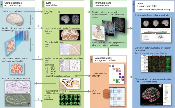
The Allen Human Brain Atlas Trends In Neurosciences. Retina and pituitary gland both share developmental origin with the brain and is also included in. The anatomical structures are all translated from the terminologia. This brain was selected for presentation in this atlas because numerous researchers. The detailed atlas of the brain in stereotaxic space is based on a brain from to the vogt collection in düsseldorf. The sectioning of the brain in the skull ensures that no significant deformation of the. The atlases of the brain in the head (macroscopic atlas) consist of serial 1cm thick sections from three human heads scanned with mri. There are several different cell types in the brain, illustrated in a specific summary page. The brain is an important organ that controls thought, memory, emotion, touch, motor skills, vision, respiration, and every process that regulates your body. Is the largest part of the brain and is composed of right and left hemispheres. Each head was sectioned either in the horizontal, coronal, or sagittal plane. High detailed anatomic coronal atlas of the human brain. There are three sections which cover: The brain atlas includes data from different species (human, pig and mouse) and different regions of the brain (bellow). They are the spatial framework for datasets such as in it provides spatial context for gene expression in the allen human brain atlas and the brainspan atlas of the developing human brain. It performs higher functions like interpreting touch, vision.
The scalable brain atlas is developed by rembrandt bakker in collaboration with many others.
This brain was selected for presentation in this atlas because numerous researchers. It performs higher functions like interpreting touch, vision. The objects, i.e., the anatomic structures, were exported as three particular sections served as reference positions. Dozens of experts have used image analysis and mathematical algorithms to evaluate the tissue sections over the years and determine the boundaries. Firstly, the human brain is divided into left and right hemispheres. The fourth edition of atlas of the human brain presents the anatomy of the brain at macroscopic and microscopic levels, featuring different aspects of brain morphology and topography. He is author and editor of several books, e.g. Building of the mri deep brain atlas. It could take a while. High detailed anatomic coronal atlas of the human brain. The brain atlas includes data from different species (human, pig and mouse) and different regions of the brain (bellow). There are three sections which cover: Like all vertebrate brains, the human brain develops from three sections known as the forebrain, midbrain and hindbrain. The brain is an important organ that controls thought, memory, emotion, touch, motor skills, vision, respiration, and every process that regulates your body. The allen mouse and human brain atlases are projects within the allen institute for brain science which seek to combine genomics with neuroanatomy by creating gene expression maps for the mouse and human brain. The atlases of the brain in the head (macroscopic atlas) consist of serial 1cm thick sections from three human heads scanned with mri. These are connected by a bundle some researchers think this area is subdivided into two or three sections. Atlas of body sections, ct and mri images ellis|ha. The template atlas of braininfo is a set of drawings of the brain that includes four cortical views and 58 coronal sections of the brain of the longtailed macaque (macaca fascicularis). He works on a digital brain atlas for planning and interindividual registration of targets in deep brain stimulation and on a spatial information j. It can be used to check the accuracy of the polygon extraction procedure (bitmap to curves). This greatly enlarged new edition provides the most detailed and accurate delineations of brain structure available. This brain was selected for presentation in this atlas because numerous researchers. The cellular structure of the brain, its anatomy, and its function. In the nissl plates cortical delineations (brodmann's areas) are provided for the first time. The detailed atlas of the brain in stereotaxic space is based on a brain from to the vogt collection in düsseldorf. The scalable brain atlas is developed by rembrandt bakker in collaboration with many others. Click to start learning with kenhub. The midbrain becomes part of the brainstem. The sectioning of the brain in the skull ensures that no significant deformation of the. The forebrain develops into the cerebrum and underlying structures;